Question 8 of 100
(demo test)
Product Demo for COA Exam Questions
-
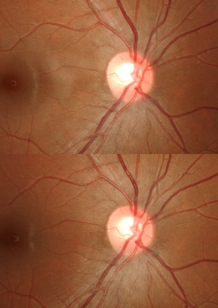
The central depression of the optic nerve is called what?
The central depression of the optic nerve is the optic cup. The round area where the nerve fibers leave the back of the eye is the optic disc, also called the optic nerve head. The central retinal artery enters the eye there and the central retinal vein leaves there. The disc is not flat. It carries a small central dip called the cup. The ring of tissue around the cup is the rim.
Nerve fibers begin at the ganglion cells on the inner surface of the retina. Their long fibers run across the retina and come together at the optic disc. Together they form the optic nerve, which carries the signal to the brain. The cup is the part of the disc where few nerve fibers pass. If fibers are lost, the cup grows larger. Staff record the size of the cup compared with the whole disc. This is the cup/disc ratio. It is written as a percentage, such as 30% cup, or as a decimal, such as 0.3 cup. An enlarging cup can be a sign of glaucoma, and the rim may thin at the same time. A pale cup and rim is called optic atrophy. The doctor may also check the disc for swelling, which is called papilledema.
Learn the back of the eye as a short list of landmarks. The disc is the nerve exit and the cup is the dip inside it. The macula and the fovea sit to the side of the disc and handle fine detail. Because no rods or cones sit on the disc, it is the physiologic blind spot. Nothing seen at that spot is sent to the brain, yet the patient never notices the gap. The fovea is the opposite: it is packed with cones and gives the sharpest vision in the eye. Naming these fundus landmarks correctly is part of the COA general medical knowledge content area.
Incorrect Answers
- A. The optic nerve head is the whole round disc, not the dip inside it. It is another name for the optic disc.
- B. The macula is a specialized area of the retina near the center of the back of the eye. It gives detailed central vision and is not part of the disc.
- D. The fovea is the center of the macula. It sits away from the optic disc and is the point of sharpest vision.
Testing Pearls
- The optic disc and the optic nerve head are two names for the same structure.
- Cup/disc ratio compares the cup to the whole disc, written as 0.3 or 30%.
- An enlarging cup with a thinning rim can be a sign of glaucoma.
- Most cone cells sit in the macula, so macular damage cuts central vision.
- The disc has no photoreceptors, which makes it the blind spot in the visual field.
References
- American Academy of Ophthalmology. Ophthalmic Medical Assisting: An Independent Study Course. Chapter 2, Anatomy and Physiology of the Eye.