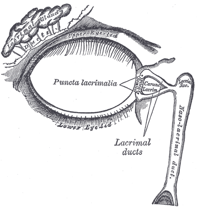
Question 60 of 100
(demo test)
Product Demo for COA Exam Questions
-
Which of the following is the correct path of the tears going from the eye to the nose?
Tears drain in this order: puncta, canaliculi, common canalicula, lacrimal sac, nasolacrimal duct. The lacrimal gland makes tears above and outside the eye. A blink spreads the tear film over the surface. Extra tears gather in a tear lake along the lower lid margin. From there they leave the eye surface through the puncta, the tiny openings on the upper and lower lid margins near the nose.
Each punctum is the doorway to a short tube called a canaliculus. There is an upper canaliculus and a lower canaliculus. These two canaliculi join together and connect to the lacrimal sac. The sac then empties through the nasolacrimal duct into the nasal cavity. The tears mix with the fluid that moistens the lining of the nose. This is why you blow your nose after crying. It is also why an eyedrop can leave a taste in the mouth. Remember that the flow goes from the lid margin toward the nose. It also goes from small tubes to a larger sac and then down the duct. Any answer that starts in the nose has the path backward.
Learn this path as one straight line: lid opening, small tube, shared tube, sac, duct, nose. A blockage in the nasolacrimal duct backs the system up and causes constant tearing. It can also inflame the sac, a condition called dacryocystitis. In a baby, the lower end of the duct may simply not be open yet. In an adult, pressing over the sac may push fluid back out through the puncta, which tells you the duct below is blocked. Staff can also press on the inner corner of the lid after a drop goes in. That step slows drainage, keeps more of the drug on the eye, and cuts how much of it reaches the rest of the body. Knowing the lacrimal drainage path is part of the COA general medical knowledge content area.
Incorrect Answers
- A. This choice puts the duct before the sac. Tears reach the lacrimal sac first and only then drop through the nasolacrimal duct.
- B. This is the path in reverse. It starts in the nose and ends at the eyelid, which is the opposite of how tears drain.
- D. This choice puts the canaliculi before the puncta. The punctum is the opening, so tears must pass through it before they enter any tube.
Testing Pearls
- Tears are made by the lacrimal gland, up and out from the eye in the orbit.
- Order to memorize: puncta, canaliculi, common canaliculus, lacrimal sac, nasolacrimal duct.
- The puncta sit on the upper and lower lid margins near the nose.
- A blocked nasolacrimal duct causes chronic tearing and can lead to dacryocystitis.
- Drops draining down this path explain the taste in the mouth after instillation.
References
- American Academy of Ophthalmology. Ophthalmic Medical Assisting: An Independent Study Course. Chapter 2, Anatomy and Physiology of the Eye.